

Small-angle neutron scattering for hydrogel structure
Recently, a group of ECRs from CeMi visited the ISIS Neutron and Muon Source at the STFC Rutherford Appleton Laboratory in Oxfordshire. The team travelled to use the neutron beam in small-angle neutron scattering (SANS) experiments to probe and investigate the structure of hydrogel matrices used to construct in vitro tissue models at the centre. Many of the projects undertaken in the centre involve the design of soft materials, of both natural and synthetic origin, with bespoke mechanical properties and bio-instructive cues to control cell interactions and behaviours. To enhance the design process and understand how these materials work it is important to evaluate their structural and mechanical characteristics. However, determining the structure of hydrogels in the hydrated state can be difficult. Techniques such as electron microscopy or micro-computed tomography (microCT) can be used to image hydrogels for analysis but require sample preparation—usually dehydration—prior to measurement or imaging, which alters the observed structure of the material. However, scattering techniques can be used to investigate the material structure in the hydrated state.

LEFT: The ISIS Neutron and Muon Source. RIGHT: The team in action analysing the scattering data.
Small-angle neutron scattering (SANS) involves exposing hydrogels to a beam of neutrons, which scatter off the atomic nuclei in the matter composing the materials. The resulting scattering profiles can be deconvoluted to reveal information about the hydrogel’s internal structure, such as their length scales, homogeneity, or structural complexity, and can be used in combination with other techniques to elucidate the mechanical properties of the materials. The team successfully prepared and measured a range of hydrogel scaffolds, and a future trip will involve simultaneous SANS and in situ rheological measurements to probe how structural characteristics of these materials change as they cross-link, swell, or are subjected to shear deformations across a range of frequencies.
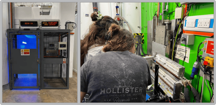
LEFT: Beam on! RIGHT: The team preparing sample cuvettes on the beam-line.