


Mechanomeds Phase 2 is an EPSRC Transformative Healthcare Technologies Second Phase funded grant.
Nowadays diagnosis is enabled by the identification of molecular markers associated with the onset of a pathological state. Nevertheless, many diseases escape this paradigm, as the biochemical fingerprint of the aberrant cells do not differ significantly from healthy ones, hindering early diagnosis and reducing the impact of treatments. One prototypical example is Leukaemia, a type of cancer that kills more than 300,000 people in the world every year. The evolution of the disease happens as we get older, but there is now evidence that cells in our body progress towards a malignant phenotype many years before they can be identified with current diagnostic techniques. This proposal will exploit mechanobiology, a field of research that has progressed in the last 10 years, as a novel method to interrogate very early changes in cellular state, bringing it closer to medical use by combining advanced biomaterials, novel microscopy techniques and robotics. Mechanobiology has taught us that cells can feel and react to their mechanical environment. For example, cancer cells are softer than normal cells. However, reorganisation of their niche causes increased tissue stiffness. Here, we will use mechanical stimulation to interrogate cells potential to become cancer cells. Cell response to these external mechanical stimuli will reveal their potential to evolve from health to disease.
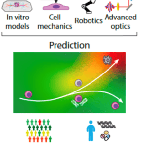
We will focus on leukaemia, a cancer that originates in the bone marrow, as normal haematopoietic stem cells, which play the essential role of making our blood, start a malignant transformation giving rise to leukemic stem cells. We have demonstrated that healthy cells and pre-malignant/malignant cells respond differently to mechanical stimulation. This project will develop an in vitro model of the bone marrow using soft hydrogels with defined mechanical and biochemical properties that host mesenchymal stem cells and hematopoietic (or leukemic) stem cells, as are found together in the marrow. We will investigate how external mechanical stimulation of the model using nanoscale vibration of controlled frequency and amplitude discriminate between healthy vs diseased systems. To monitor these mechanical changes in the in vitro model we will use Brillouin microscopy in a biological context. This technique is based on the propagation of acoustic waves in the system to characterise mechanical properties and will allow detailed mapping of stiffness of the bone marrow model as a function of time – importantly in a non-invasive way. Moreover, the level of mechanical stimulation will be dependent on the readout provided by Brillouin microscopy that will feed into a control system to alter the level of the mechanical vibrational stimulation imposed on the bone marrow model. We will develop the technology to have a robust on-chip system that includes the bone marrow model and integrates mechanical stimulation.
We will use the technology in two clinical applications:
(1) to assess whether the technology can predict leukaemia which can be induced as an off-target effect of the treatment (chemotherapy/radiotherapy) of solid tumours and
(2) to assess whether the technology can predict malignant transformations in heaematopoeitic stem cells that happens with age, eventually leading to leukaemia.


The University of Glasgow / University of Strathclyde
Centre for the
Cellular Microenvironment
Cellular Microenvironment